Author: Milicent Cranor

  • Biological Map in JFK’s Neck Points to South Knoll

    Biological Map in JFK’s Neck Points to South Knoll


    If you want to know where one of the shots that hit John Kennedy came from, you can consult a biological map, and the one in Kennedy’s neck is pretty reliable. Other researchers present bullet paths through the head, but the one in the neck is easier to see.

    If the wound in Kennedy’s throat was an entrance, this of course means that one shooter was firing from in front of the motorcade. (Please go here to see some reasons to believe it was an entrance.) But the path through Kennedy’s neck can tell you approximately where in front the shooter was. Consider the three dots below that represent damage reported by Parkland Hospital:

    • A small hole in the skin in the middle of the neck
    • A larger hole in the right side of his trachea
    • Bleeding in the area of the right mediastinum over the lung

    Connect these three dots and you have a diagonal line across Elm Street that leads to an area in front—and to the left of Kennedy. The south knoll.

    In other words, the damage in the neck shows the path of a bullet going northeast—which means it came from the southwest.


    No Fourth Dot

    If the wound in Kennedy’s throat was an exit instead of an entrance, then we would have to assume that a bullet entered the back and exited the throat. But in an article published long ago, the late John Nichols, MD, PhD explained why this could not have happened: the wound was only 5 centimeters or 1.9 inches from the midline of the back and even closer to outer edge of the spine itself. In its hypothetical journey to the middle of the throat, the bullet would have to go through the spine.

    And he showed that for the bullet to avoid hitting the spine, it would have to have entered the back further to the right than it actually did.

    Here is a scan showing the cross-section of the neck of a man whose size was very similar to Kennedy’s. It was done by David Mantik, MD, PhD to demonstrate what Nichols was talking about.

    The red line begins in the lower right-hand corner—where the bullet wound was in Kennedy’s back. The wound could not be probed and seemed to stop within an inch after penetrating. In any case, a medium-high velocity bullet could not have followed the path below without creating tremendous, obvious damage, shattering the bones it went through as well as damaging tissue lateral to its path.

    mantik scan

    X-rays of Kennedy’s spine showed no such damage. The shadow of a line appeared between two parts of the spine, suggesting a separation of the transverse process and where it attaches. Promoters of the Lone Nut Theory have tried to use it as proof of a shot at the seventh cervical level—but buried in the HSCA Hearings is an expert radiologist’s report of several additional shadows, proving they are all meaningless artifacts:

    Unenhanced x-ray: “The first rib appeared to be separated from the sternum …” (JFK Exhibit F-34). Enhanced x-ray: “there appear to be fractures of the posterior aspects of the 2nd, 3rd, and 4th ribs. These are artifacts.” (7 HSCA 219)  (Please go here to see my story on this, and more.)

    The lead pathologist who performed the autopsy, James Humes MD, testified that they saw no fractures in the vertical column, that is, the spine. His words:

    … we examined carefully the bony structures in this vicinity [of the back wound] as well as the X-rays… and we saw no such evidence, that is no fracture of the bones of the shoulder girdle, or of the vertical column, and no metallic fragments were detectable by X-ray. [I believe “vertical” should have been “vertebral,” possibly a transcription error.]


    Reversing the Path

    Any bullet—whether from the front or the back—missed all parts of the spine. And so I propose we use, as a biological map, Nichols’s diagram of a path that misses the spine:

    nichols

    Where the bullet stopped, we do not know. Nor do we know what happened to it. It is entirely possible that it was found during the autopsy and furtively removed by one of the pathologists, his back to the audience, his body obscuring Kennedy’s.

    Trusting the Parkland Hospital doctors’ report—but not the pathologists’—we do know the damage suggests the bullet went to the right somewhere inside Kennedy, just above the right lung. And so I suggest we use the uppermost path in Nichols’s diagram as our map. It goes far enough to the right to miss the spine.

    Below is another diagram from the Nichols article which I have cut to include only the path through the front of the body. In other words, I have disconnected it from the back wound. It shows a simple overhead drawing of Kennedy and his right arm:

    angle schematic

    You will have to orient this diagram in relation to where the limousine was at the time of the strike, and it may be impossible to get it exactly right. I suggest finding several possible locations, bracketing them—no earlier than this position, no later than that position.

    I would wish you happy hunting, but this is really a grim business. 

  • Ricochet of a Lie

    Ricochet of a Lie


    Ricochet of a Lie

    And Kennedy’s Throat Wound

    (A version of this essay was previously published under the title “The Magic Throat.”)


    If you believe the gaping hole in the front of Kennedy’s neck was the result of body alteration by unknown persons, the facts below may change your mind.


    Commander James Humes, M.D., the lead pathologist who performed the autopsy on President John F. Kennedy, was interviewed in 1992 by the Journal of the American Medical Association (JAMA).1 Once again, he and his team were going to set the record straight.

    As often happens on these occasions, Humes provided a fresh crop of contradictions.   One such contradiction is the subject of this article: the size Humes gave JAMA for the incision in Kennedy’s throat: less than half the size he reported earlier,2 3 and less than half the size as it appears in autopsy photographs.  

    What intrigues me about this new size is its striking similarity to the one described by Malcolm Perry, M.D., to David Lifton, author of “Best Evidence.”4


    1964, James Humes: Large Incision

    According to Humes’s autopsy report,5 the tracheotomy incision was 6.5 centimeters, but Humes told the Warren Commission it was between 7 and 8 centimeters.6 The latter description seems to match what shows in autopsy photographs. (Note: “tracheotomy” is sometimes spelled “tracheostomy”; both are correct.)


    1980, Malcolm Perry: Small Incision

    The best selling Best Evidence by David Lifton was published in 1980. In it, Lifton quotes Malcolm Perry as saying the incision was only 2 to 3 centimeters wide. When Lifton asked if it could have been 3.5 centimeters, Perry agreed. Could it have been 4 centimeters? Perry “hesitated a bit, then said yes.” How about 4.5 centimeters? Perry said he “really doubted it was that large. It just wasn’t necessary.” Lifton added, “Perry sounded distinctly uneasy.” Lifton quoted other Parkland doctors who corroborated Perry’s claims.7

    Perry’s first answer, “2 to 3 centimeters wide,” is consistent with the routine tracheotomy incision as diagrammed in Lifton’s book.

    Not the kind of incision made on a victim of a penetrating neck trauma.


    1992, James Humes: Small Incision

    Humes told JAMA that “The tracheostomy was a gaping wound, about 3 to 4 centimeters around ….”8

    Only 3 to 4 centimeters “around”? This is how one describes a perimeter. Translated, this would make the incision across no more than 2 centimeters in horizontal width—about the size Malcolm Perry gave David Lifton. A very interesting match indeed.


    What Did Malcolm Perry Actually Do?

    Small incisions are created for routine tracheotomies—the kind performed for patients with severe breathing problems, such as those due to pneumonia, for instance. David Lifton investigated and then publicized the appearance of a routine tracheotomy incision.

    What David Lifton did not investigate at all, apparently, was this relevant question:

    How do emergency room doctors respond to a penetrating wound in the neck, regardless of whether the patient has wounds elsewhere in the body?

    Doctors must quickly find and repair any life-threatening damage that has been done—and that means creating a fairly wide incision, one large enough to visualize the carotid arteries, for example. (These arteries are not in the middle of the throat; they are off to either side of the neck.)

    From Perry’s and Humes’s descriptions, it sounds like Kennedy had a “collar incision,” similar to the one shown below from the Journal of Trauma 1978; 18 (1) : 2-77 (see the Addendum at the end of this article for more extensive documentation).

    incision

    In Kennedy’s case, it was already clear that he was having trouble breathing, and one doctor placed an endotracheal tube which proved to be inadequate. So when Perry arrived, he performed a tracheotomy. And, he said, he “made an incision right through this wound which was present in the neck.”9

    But that was not all that Perry did. He made an incision large enough to allow him to inspect the carotid arteries, the mediastinum, and other structures.

    “Once the transverse incision through the skin and subcutaneous tissues was made, it was necessary to separate the strap muscles covering the anterior muscles of the windpipe and thyroid. At that point, the trachea was noted to be deviated slightly to the left and I found it necessary to sever the exterior strap muscles on the other side to reach the trachea.”10 (His testimony on another day is similar.11)

    “I also made it big enough that I could look to either side of the trachea … I didn’t know whether I was going to encounter carotid arteries or whatever. But the path of the bullet clearly put those vessels at risk as well as the trachea, so I made the wound big enough to do that … How big it [the incision] was, I don’t know … . I made it big enough to control an underlying bleeding blood vessel if necessary.”12

    Please see the Addendum for quotations from articles on this subject—including some from a chapter in a textbook written by Malcolm Perry himself.


    Why Perry Described Such a Small Incision: A Theory

    Malcolm Perry was aware of Humes’s sworn testimony that, on the night of the autopsy, he knew nothing about a bullet wound in the throat, that he thought only a tracheotomy had been done.

    Caught off guard by Lifton’s question, Perry probably thought he had better not say anything that would contradict Humes’s story. 

    Perry was apparently confident that Humes’s earlier report of a large incision was not well known. He has said more than once that he never met anyone who had read the Warren Commission Hearings.


    Why Humes Revised the Size: A Theory

    Small incisions are not done in the presence of a penetrating wound in the throat.

    If Kennedy only had a small opening in his throat, this would have made it easier to believe Humes’s story of thinking the president only had a tracheotomy. This may have been the reason Humes gave such a different description of the incision to JAMA.

    Furthermore, he may have worried that, had he described a wider incision, one that was more consistent with those performed in the presence of trauma, then one of JAMA’s readers might well have asked, how could he have been so sure the only abnormality in JFK’s throat was the remains of a tracheotomy? Especially in the presence of a bullet wound in the back?


    What May Lie Behind the Revisions

    Had Humes admitted to being aware of a throat trauma, he would have then been obliged to deal with it: dissect its path, obtain tissue specimens, document its characteristics, photograph it close-up and under magnification.

    What if a proper study of the wound showed it to be an entrance? This would mean that at least one shooter was in front of Kennedy. But Humes and his team were under a great deal of pressure to prove the president’s wounds were created by one sniper, located above and behind.

    Better to feign ignorance of a wound that may contradict that conclusion. Better to wait until after the body was gone, when it was too late to document its nature—entrance or exit—and its alleged connection with the wound in the back.13


    But Humes Did Know

    It seems that, even on the night of the autopsy, Humes et al. did know that a bullet had passed through Kennedy’s throat, either coming or going.

    The bruising in the neck is what told him, bruising that could only have occurred during the motorcade—when the heart was still beating normally. What’s more, possibly out of vanity (and to avoid looking stupid to his peers), he wanted the Warren Commissioners to know that he knew, that he had not missed something so important, or its significance.

    “When examining the wounds in the base of the President’s neck anteriorly … we noted, and we noted in our record, some contusion and bruising of the muscles of the neck of the Present. We noted that at the time of the postmortem examination.”14

    He went on to describe wounds that were created by the Parkland doctors: incisions made in the chest for the placement of chest tubes, and in the arm and ankle for administering intravenous fluids—wounds that did not lead to bleeding and bruising, because they were made later, when the heart was barely beating.

    “Those wounds [made at Parkland, on the chest and in the arm and ankle] showed no evidence of bruising or contusion … which made us reach the conclusion that they were performed during the agonal moments of the late President … when the circulation was … seriously embarrassed, if not nonfunctional. So that these wounds … were performed about the same time as the tracheotomy wound …”15

    “So, therefore, we reached the conclusion that the damage to those muscles on the anterior neck just below this wound were received at approximately the same time that the wound here on the top of the pleural cavity was, while the President still lived and while his heart and lungs were operating in such a fashion to permit him to have a bruise in the vicinity … so we feel that, had this missile not made its path in that fashion, the wound made by Doctor Perry in the neck would not have been able to produce, wouldn’t have been able to produce, these contusions of the musculature of the neck.”16

    Ignoring Humes’s comments above, Commissioner Allen Dulles then completely changed the subject.17

    In later years, J. Thornton Boswell, the pathologist who assisted Humes, actually described seeing, on that same night, the remains of the bullet hole itself, “part of the perimeter of a bullet wound in the anterior neck”,18 and they concluded that night that the bullet exited the throat.19 When talking to the ARRB in 1996, Humes denied seeing the remains of the bullet wound, or knowing anything about its existence.20

    The lead photographer, John Stringer, described the pathologists putting their fingers into the gaping hole trying to feel for anything sharp, looking for bullet fragments, in other words.21 John H. Ebersole, M.D., Acting Chief of Radiology, said that Humes had been in touch with the Parkland doctors that same night, and, clearly, had been informed of the bullet wound in the throat.22 (This is not a comprehensive treatment of the subject; there is yet other testimony relative to this issue not included here.)


    Comic Relief: Humes “Explains”

    “And we wondered, where’s the bullet? You know. Should have called Dallas right then and there. It would have saved me a lot of worry and grief for several hours, because x-rays hadn’t found it for us. Like it could have been in his thigh or it could have been in his buttock. It could have been any damn place …”23

    So, it could have gone any damn place—except a few inches away out the hole in the front?

    “We didn’t know where it went. It was obvious after we talked to the doctors the next morning where it went. It went out. That’s why we couldn’t find it. And we weren’t going to spend the rest of the night there, you know. Meantime, George Burkley is telling me, you know, the family wants to get out of here sometime tonight. Then we proceeded with the dissection of the lungs, heart and abdominal contents and so forth.”24

    Note: Kennedy was not shot in the abdomen.

    “… it wouldn’t make a great deal of sense to go slashing open the neck. What would we learn? Nothing you know.”25

    Have sympathy for the late James Humes. He not only had to cover his own ass, he had to cover the collective ass of those who were pressuring him to come up with the “right” answers. He never did seem sure of the script.


    Was it an Entrance Wound?

    We have no way of knowing what the nature of Kennedy’s throat wound was but, considering all the lies told about it, Humes et al. probably thought it was an entrance. Based on the wound’s characteristics as reported, nothing seems to justify declaring it an entrance or an exit although, grossly, it was most typical of an entrance.

    • Exit wounds can be small;
    • Ragged wounds can be entrances;
    • Entrance wounds need not have abrasion collars, but often do;
    • Shored exit wounds always have pronounced abrasion collars.
    • Non-shored exit wounds do not have abrasion collars.

    One thing does seem certain: if the wound had been an exit, the bullet had to have exited above the shirt; it could not have gone through the shirt (the bottom part of the collar band), as alleged. This would have created a “shored wound.” In shored wounds, an obvious abrasion collar is created26 with little bits of skin hanging (like the flakes of a sunburn) as the shoring material—which usually sticks to the wound—is pulled off the victim.27

    Furthermore, skin would have been left behind on the shirt: upon exiting, the bullet would have crushed the skin against the shirt on its way out, skin that would have been visible to the naked eye.28 The FBI would surely have loudly announced such evidence had they found any.

    Kennedy’s throat wound did not have the wide, pronounced abrasion collar of a shored exit wound, but it apparently did have the abrasion collar of an entrance wound.29

    As mentioned above, exit wounds can be small, but would a jacketed bullet travelling at medium high velocity through the loose, wrinkled, tanned, leathery skin of Kennedy’s neck leave a small wound?

    Or—unhindered by any shoring material—would the bullet pull the skin forward on its way out, tenting the skin, creating the lacerations of a typical “stellate” wound of exit?

    (All of the small exit wounds in the photos that I could find were in what looked like taut, young skin in areas not unexposed to the sun and not subjected to a lot of stretching, and wrinkle-causing movement, like the neck and wrists.)

    For more on the subject of bullet wounds (including photographs and additional references) please see my article, Trajectory of a Lie Part I. The Palindrome” at www.history-matters.com.


    Conclusion

    Commander James Humes testified that, on the night of the autopsy, the only abnormality seen in Kennedy’s throat was a tracheotomy incision. For the following reasons, this seems to have been a cover story:

    • Context: there was an entrance wound, only inches away, in the back;
    • The incision was larger than one for a typical tracheotomy, though, by itself, this is not reason to suspect trauma;
    • The strap muscles were severed; this is not done in routine tracheotomies;
    • Humes testified he saw bruising in the throat muscles that he said he knew could only have occurred during the motorcade, when the president’s heart was still functioning normally;
    • Other doctors testified that they all knew of the bullet wound in the throat;
    • Part of the perimeter of the hole created by the bullet was seen.

    By denying awareness of the wound, Humes avoided documenting its nature. (He also avoided properly documenting the back and head wounds, all for the same reason apparently.)

    A few years later, in an apparent effort to support Humes’s story, the surgeon who performed the incision, Malcolm Perry, M.D., described it as small enough to be typical. This same surgeon was most accommodating when it came to revising other trajectory-related details.   Nearly 30 years later, Commander Humes himself reported the incision as small, radically contradicting his own testimony to provide a description that just happened to agree with Malcolm Perry’s.

    It is also possible that each doctor simply had a malfunction of memory.


    ADDENDUM

    Standard Response to Penetrating Wounds of the Neck

    What was the standard emergency response to a penetrating injury of the throat—whether or not the trachea (windpipe) is injured, and whether or not there is another life-threatening wound?

    Exploratory surgery—however quick and limited—was and is the standard. The incision Perry made was apparently similar to the one shown in the diagram below:30

    incision

    According to the 1971 issue of the American Journal of Surgery, the performance of an “exploratory” in these circumstances had been standard since World War II:

    “The general surgical principle of mandatory exploration of all penetrating wounds has become established during the past three decades.” 31

    Parkland Policy on Penetrating Neck Wounds

    “Since 1957 it has been the policy at Parkland Memorial Hospital to explore virtually all penetrating wounds of the neck regardless of the clinical impression as to the severity of the injury …”

    “The incision was planned to allow full exposure of the tract of injury. Proximal and distal control of the major vessels was also considered in the length and position of the incision. The sternocleidomastoid (“strap”) muscle and/or other neck muscles were taken off the insertion or transected whenever necessary to provide adequate exposure.”32

    The textbook Principles of Trauma Care contains a chapter written by Malcolm Perry, “Penetrating Wounds of the Neck.”33 Perry’s list of findings that indicate the need for exploration include some that applied to Kennedy:

    • Bleeding
    • Large hematoma
    • Shock
    • Respiratory distress
    • Subcutaneous emphysema
    • Blood in the oropharynx

    Finally, an exploratory is even appropriate in the absence of these signs because life-threatening damage can hide behind a superficial looking wound. The authors of the 1971 paper discovered the following:

    “… one third of the patients with injury to a major structure had no clinical evidence for this. This latter group included one patient with a through and through bullet wound of the carotid artery.”34


    Notes

    1 D. L. Breo, “JFK’s death-the plain truth from the MD’s who did the autopsy.” Journal of the American Medical Association 1992; 267: 2794-2803, here p. 2798.

    2 Autopsy Report. MD3, ARRB Master Set of Exhibits.

    3 J. J. Humes, Warren Commission Hearings, Vol. II, p. 361 (“some 7 or 8 cm in length”).

    4 D. S. Lifton, Best Evidence. Carroll & Graff, 1980, p. 238.

    5 Autopsy Report.

    6 WCH II, p. 361.

    7 Lifton, p. 238.

    8 Breo, p. 2798.

    9 M. Perry, Warren Commission Hearings, Vol. VI, p. 10.

    10 WCH VI, p. 10.

    11 Perry, Warren Commission Hearings, Vol. III, p. 370.

    12 Perry, ARRB Testimony, August 17, 1998, pp. 24-25.

    13 Humes, HSCA Testimony, September 7, 1978, p. 330.

    14 WCH II, p. 367.

    15 WCH II, p. 367.

    16 WCH II, p. 368.

    17 WCH II, p. 368.

    18 J. T. Boswell, HSCA Testimony, August 7, 1977, pp. 8, 12.

    19 Boswell, ARRB Testimony, February 26, 1996, pp. 34, 45.

    20 Humes, ARRB Testimony, February 13, 1996, p. 115.

    21 J. Stringer, ARRB Testimony, July16, 1996, pp. 191-2.

    22 J. H. Ebersole, HSCA Testimony, 1978, p. 20; pp. 51-52.

    23 Humes, ARRB Testimony, p. 113.

    24 Humes, ARRB Testimony, p. 113.

    25 Humes, ARRB Testimony, p. 36.

    26 V. J. M. Di Maio, Gunshot Wounds, Practical Aspects of Firearms, Ballistics, and Forensic Techniques, Second Edition. CRC Press, 1999.

    27 J. C. Aguilar, “Shored gunshot wounds of exit.” American Journal of Forensic Medicine and Pathology 1983; 4(3): 199-204.

    28 Aguilar, “Shored gunshot wounds of exit.”

    29 M. Perry, HSCA p. 302; C. Baxter, Warren Commission Hearings, Vol. VI, p. 42.

    30 For the diagram: J. D. Saletta, R. J. Lowe, L. T. Lim, J. Thornton, S. Delk, and G. S. Moss, “Penetrating trauma of the neck.” Journal of Trauma and Acute Care Surgery 1976; 16(7): 579-587; D. C. Blass, E. C. James, R. J. Reed, C. W. Fedde, and A. L. Watne, “Penetrating wounds of the neck and upper thorax.” Journal of Trauma and Acute Care Surgery 1978; 18(1): 2-7.

    31 “Exploratory since WW II”: C. Ashworth, L. F. Williams, and J. J. Byrne, “Penetrating wounds of the neck. Re-emphasis of the need for prompt exploration.” American Journal of Surgery 1971; 121: 387-391.

    32 Parkland: R. F. Jones, J. C. Terrell, and K. E. Salyer, “Penetrating wounds of the neck: an analysis of 274 cases.” Journal of Trauma and Acute Care Surgery 1967; 7(2): 228-237.

    33 M. Perry, Chapter 16, “Penetrating Wounds of the Neck.” In: G. T. Shires, ed., Principles of Trauma Care, Third Edition. McGraw-Hill Book Company, 1985.

    34 Ashworth, Williams, Byrne, “Penetrating wounds of the neck.”

  • The Magic Scalp

    The Magic Scalp


    Witnesses who saw JFK’s head up close after he was shot, describe damage that is quite different from what shows in certain autopsy photographs and x-rays. And the contrast between the two – the damage they describe, and the evidence on films is so radically different, many researchers suspect evidence tampering.

    There are people who defend the authenticity of the evidence by “explaining” the problem with theories that may sound reasonable – but some of these people promote their work in the following ways: (a) they omit significant information that challenges their ideas; (b) they pad their work with irrelevant information – thus obscuring the paucity of proof of their main thesis; (c) they try to shape ambiguous language to mean only what they want it to mean; (d) they make amateurishly omniscient assertions… “This is irrefutable proof… There’s no other explanation… This has to mean…”; (e) they list people who presumably agree with them without showing the reader what exactly they had agreed with, and some of the people are in rest homes, or in graves, or otherwise are hard to reach.


    JOHN CANAL’S THEORY

    John Canal, retired USAF Senior Master Sergeant, has a theory designed to explain away two major issues with JFK’s head wound: (1) how the alleged entrance wound, described by the pathologists as low in the head, was four inches higher, as interpreted by medical panels who later studied the photos and x-rays (not the body); (2) why the back of the head pictures show no wound at all, not the big obvious opening described by Parkland doctors and others, and not the smaller entrance wound.

    Canal has promoted his explanation in three different articles, one in 2013 in Max Holland’s Washington Decoded, and two more in something called “Student Operated Press,” The SOP 2014, and The SOP 2015.

    Canal’s theory has multiple parts: (a) he insists the main photo in question (see below) was taken after the morticians reconstructed the head; (b) reconstruction involved moving the scalp from the back to the front of the head, to cover any gaps that might be seen, should there be an open casket funeral; (c) the entrance wound, low in the back of the head, got dragged to the top of the head; (d) the back of the head looks undamaged because the photo was taken after bone was put back in and the scalp was sewn shut.

    In the SOP in 2015, he asserts:

    “Again, because the BOH [back of head] photographs show the entry wound in the scalp high in the cowlick, the fact that the skull entry was low (approximately two inches above the hairline) is incontrovertible evidence the BOH photographs were taken after the autopsy when reconstruction of the BOH by the morticians was completed.”

    This reconstruction is supposed to hide damage from viewers of an open coffin funeral? Then why did they leave that bone flap in front of the ear still flapping away? Surely this photo was not taken when reconstruction was completed. We can be generous and say it was taken during the reconstruction, but Canal presents no proof of that either.


    JOHN CANAL’S “PROOF”

    Before we go any further, let’s take a good look at the photo in question:

    photo

    (People say this photo shows no damage, other than the small white image near the hairline, said to be adherent brain tissue. Yet just above it is an odd, light-colored, angular formation – but that is a side issue not relevant to this essay.)

    What is presumed to be the wound is a flat-looking area of light, watery, reddish brown – one of many in the photo – with hairs growing out of it, apparently. If you blow it up, you will see an “X” crisscrossing through it. Possibly these are hairs.

     blowup

    “X” marks the spot. Hairs?

    If you have ever seen an entrance wound in scalp created by a jacketed bullet traveling at medium high velocity, you will know that it looks nothing like this. The skin is crushed between bullet and bone, and appears quite dark. Lift up the scalp, and the edges of the hole are still apparent. The X’ed image in the photo does not look like a hole at all.

    But this is how the wound is described on page 104 of HSCA Volume VII: “The inferior margin of this wound, from 3 to 10 o’clock, is surrounded by a crescent-shaped reddish-black area of denudation, again presenting the appearance of an abrasion collar, resulting from the rubbing of the skin by the bullet at the time of penetration. From 12 to 3 o’clock, there is a suggesting of undermining, that is, tunneling of the tissue between the skin surface and the skull…” They put this description under a drawing of the photograph.

    The above description is a conflation of the drawing and photograph. I see no “reddish-black area of denudation” in the photograph – but the drawing certainly has a black and white equivalent.

    Now please look at what is supposed to be an accurate drawing of this photo:

    dox

    As you can see, the “wound” is much darker, has more dimension, and rolled edges. It looks like a hole at least. Not one of these characteristics appears in the photograph.

    In his beautifully written essay on the medical evidence, Gary Aguilar, MD also compared the two, and said of the drawing, “the small spot visible just to the right of the top of the ruler is exaggerated in this diagram. It is significantly smaller in the original photograph.” (And there are probably other researchers who have described the contrast between the photo and the drawing.)

    It is the drawing, not the photo, that John Canal presents in his articles. To the uninitiated, the drawing may appear to display a wound.

    Hair Surrounding “Wound” Too Long for EOP Area

    Kennedy’s hair was quite short in the back, including the area where a bullet allegedly entered. And it was even shorter below that area as it approached the neck. As you can see from the photo, the hair all around the “wound” in the cowlick is much too long.


    WITNESSES DO NOT SUPPORT CANAL’S THEORY

    No witnesses said pictures were taken during or after the reconstruction of the skull. None said scalp from the EOP area was dragged to the top of the head.


    JOHN STRINGER

    Canal’s star witness – John Stringer – who Canal insists took photographs of the reconstructed skull after the autopsy, said in 1996:

    “We took no photos during or after the embalming.”   (ARRB 4/8/96, p.5)

    And from his longer deposition, it seems clear that Stringer only took photos during the autopsy – including the ones John Canal claims were taken after reconstruction of the skull. (ARRB 7/16/96)

    But in his Washington Decoded article, Canal gives prominence to what he seems to have gotten Stringer to say in 2011, when Stringer, who was born in 1918, said at the age of 93:

    “In one statement, Stringer wrote, ‘I may have taken some pictures after midnight, but I just can’t remember, it’s been too long.’ [April 30, 2011] In view of Stringer’s 1996 testimony that he did not arrive at his home (not far from the morgue) until about 4 AM, however, and that cosmetic reconstruction of the head began shortly after 11 PM, the inference that he took pictures later as well as earlier is reasonable. It is also consistent with a statement in his book MEDPHOTO that he took photos at various times throughout the procedure and whenever he was directed to do so. [21]

    Buried in Reference 21 is Stringer’s earlier statement to the ARRB:

    “[21] John Stringer, MEDPHOTO: Snapshots of Life in Peace & War with the US Navy (Mooresville, NC: Wishbone Creative Product Services, 2008), 37. In an ARRB interview, Stringer also said that, ‘We took no photos during or after the embalming.’ In contrast, before that statement, he stated photos were taken “throughout the autopsy.”

    Notice Canal’s last remark – as if taking pictures “throughout the autopsy” is supposed to suggest he took them after the autopsy. He pads his article with quotes from a number of people using those words, “throughout the autopsy”, as if that meant afterwards. He also conflates “late photography” to mean after the autopsy:

    “Other witnesses at the postmortem whose observations support late photography [emphasis added] included Captain John Stover, an officer at the Bethesda Naval Medical Center; John Van Hoesen, one of the morticians; Joseph Hagan, supervisor of the team of morticians; Floyd Riebe, the assistant autopsy photographer; Jan Rudnicki, who assisted the autopsy doctors; General Godfrey McHugh, who observed the autopsy; Jerrol Custer, an X-ray technician; and James Sibert, one of the two FBI agents who observed the autopsy.”

    Notice that he leaves out Tom Robinson, the mortician who said many things that challenged the official story.  

    TOM ROBINSON (mortician, not mentioned by Canal):

    “When asked, Mr. Robinson said he had no recollection of photography the night of the autopsy, one way or the other – no recollection whatsoever.” (ARRB 6/21/96, p.3)

    JOSEPH E. HAGAN (mortician):

    “He does not recall, one way or another, whether any photographs were taken during Gawler’s work on the President’s body.” (ARRB 6/11/96 p.4)

    Before the morticians worked on the body,

    “Hagan said that when he arrived… the autopsy was almost over; he only had to wait in the gallery about 20 minutes before the autopsy was concluded. The body of the president was being ‘cleaned up.’ Hagen said photos were being taken, but could remember no details…” (ARRB 6/11/96, p.3)

    JOHN VAN HOESEN (mortician):

    Canal presents this passage from David Lifton’s book that he (Canal) says suggests photos were taken after head reconstruction. But he seems to be describing nothing more than having to wait on the autopsy and the picture-taking before they could begin their own work. And his comments seem to echo Hagen’s (see above):

    “Van Hoesen: When we got up there, nothing had been started; then we had to wait for the autopsy; and then periodically, more pictures were being taken, “you know, different angles and so forth; where the entry was, and so forth; this angle, and that angle …  Lifton, Best Evidence, 666.”

    Canal does not report what Van Hoesen told the ARRB, and it does not help his theory any.

    “He could not remember, one way or another, whether photographs were taken during the embalming and reconstruction process.” (ARRB 9/26/96, p.3)


    LOGIC DOES NOT SUPPORT CANAL’S THEORY

    Scalp Borrowed From an Area Missing Scalp?

    It is well-established that witnesses, including a prominent brain surgeon at Parkland Hospital, said both bone and scalp were missing from an area in the back of the head that included the occiput. The lead pathologist who wrote the autopsy report, James Humes, was vague about a lot of things, including how much of the great defect involved occipital bone, but he did admit the wound was “somewhat” in that area. In any case, the back of head photograph presented earlier in this essay shows no such defect in bone or scalp.

    According to Canal, scalp was borrowed from that area – even though it was already missing scalp – to cover the top right of the head.

    But Canal ignores the testimony about the large hole in the scalp – which he describes as merely “torn.”

    “Specifically, the cosmetic repair involved first suturing the tear in his rear scalp until it was closed, and then, after undermining, stretching the scalp until it covered the large deficit in the top/right/front of Kennedy’s head where the bullet had exited, bone was blown out, and scalp missing or badly damaged.

    “The stretching of the scalp occurred after the autopsy was completed, sometime around 11 PM, and once the embalming and cosmetic restoration of the body commenced. The morticians had only the best of intentions when they took advantage of the fact that the rear scalp had only been torn, and was both repairable and useful for another purpose. They were simply trying to cover that large deficit in the head in anticipation of an open-casket funeral.” (Washington Decoded)

    “The BOH opening, in all likelihood, was created after the bullet’s explosive impact exposed the president’s brain through a tear in the rear scalp and an opening between two or more dislodged (but not blown-out or missing) pieces of loose rear skull. This observation is supported by the fact that the lateral X-ray shows no missing rear bone whatsoever. Dr. J. Thornton Boswell, one of the prosectors, did say in 1996 that he repositioned some bone pieces before the X-rays and photos were taken; it seems logical that he pushed some loose pieces of skull (dislodged but still adhering to the scalp) roughly back into place. (Washington Decoded)


    SUMMARY

    1. Not one witness mentioned by Canal says that photos were taken after the autopsy.
    2. In the photograph, there is no proof of a small entrance wound. But Canal does not show the photograph. He shows the drawing of it, even though the drawing, and the HSCA description of the wound, do not match the photo.
    3. What Canal says is the wound imported from just above the EOP is surrounded by hair that is much too long for that area.
    4. How Canal avoids the problem that scalp would not likely be borrowed from an area that has a sizable hole in it: he claims falsely that in the back of the head, the scalp was merely torn.

    Despite the absence of proof in any of his articles, he said “The evidence for these BOH photographs being taken after the autopsy is irrefutable and so extensive it would not be practical to list here.” The SOP 2015.

    What is “extensive” is the list of problems in Canal’s essays, but I focused only on those that seem to be the worst.

    I wrote this essay in response to an email I received from a student at a college in Texas. She was having trouble making sense of these articles and someone referred her to me.


    ADDENDUM

    I have no opinion as to whether photographs were taken of a reconstructed skull, or when. I only know that witnesses do not support such a claim. While their testimony may be inaccurate or even false, it should be presented. When an author publishes a theory, the author should be the first to let the reader know whatever challenges that theory.

    The testimony of Tom Robinson (excerpted below) contradicts John Canal’s assertions. Canal said the wound in the back of the head consisted only of “torn” scalp and, rather than an area of missing bone, that bone was merely displaced. He also said the bone was put back during the autopsy. But all the pathologists did was to replace loose bone that had fallen out during their probing. But they could not replace bone that was missing in the first place.

    When the body was turned over to the morticians, the skull was missing a large area of bone in the back. Its appearance was not the problem; it would not have been visible during an open casket viewing. But it had to be closed to prevent embalming fluid from leaking through it.

    Robinson did not describe working on the top and side of the head, so we have no details about what was done in these areas – the only parts that would show in an open casket. He just said the top appeared to be “all broken” but not open like the wound in the back.

    When shown the back of head photo, he said the wound was just above the white spot in the hairline. If he was right, this would mean the wound was rather low.

    This is how he described the reconstruction of the area of missing bone in the back:

    “Robinson said that Ed Stroble… had cut out a piece of rubber to cover the open wound in the back of the head… the piece of rubber was slightly larger than the hole… the rubber sheet was a circular patch about the size of a large orange… He said the cranium was packed with material during reconstruction… The rubber sheet was used outside of this material to close the wound in the area of missing bone. The scalp was sutured together, and also onto the rubber sheet to the maximum extent possible and the damage in the back of the head was obscured by the pillow in the casket…”


    Location of Hole in Back; Condition of Top of Head

    robinsonA


    Size of Area of Missing Bone, Reconstruction

    robinsonB

  • John Lattimer Never Quit: The Thorburn Business


    (A longer version of this article was published several years ago.)


    The location of Kennedy’s back wound is a major obstacle for the Single Bullet Theory (SBT). It is the first of several dots that needed to be connected – all at a downward angle: (1) the alleged sniper’s nest, (2) the back wound, (3) the throat wound, (4) Governor’s Connally’s back wound (the exit in the front is not questioned), and (5) his wrist wound.

    Shall I burden the SBT with yet one more dot? (6) The little fragment that ended up in Connally’s thigh not far beneath the skin – and the mysteriously large, 10 mm, round, corresponding hole in his pant leg.

    The back wound was just too low. If the bullet actually went through JFK’s torso and exited his throat – it would have done so at an upward angle. For that bullet to have reached Connally’s pants, he would need to have lifted his leg rather high in the air, above his seat.

    I doubt that even John Lattimer could have found a way to show that on the Zapruder film. (But who knows what went on when the president’s car went behind the Stemmons sign? Maybe Dale Myers could do another study, proving that Connally did raise his leg high above the seat while the car was out of sight.)

    Lattimer found a way to raise Kennedy’s back wound, up, up, up – all the way to the sixth cervical vertebra (C-6), a tremendous feat.

    In 1977, he published a paper (1) in which he claimed the movements of Kennedy’s arms and hands – he seems to be trying to grab his throat – are symptoms of damage at the C-6 level. To give this paper more authority, he put the names of two other prominent doctors on it: Edward Schlesinger, a neurosurgeon, and H. Houston Merritt, a neurologist. He wrote:

    “The precision with which the signs of a spinal cord lesion fit with the other evidence (loose fragments) that the tip of the transverse process of C-6 (not C-5 or C-7) had been struck by the bullet … ” (p. 287)

    To prove it, the authors cited a nineteenth century paper (2) on a patient treated by neurologist William Thorburn, a patient who fell backwards, and was found with his arms in a strange, unnatural position, a position that was, presumably, similar to Kennedy’s. The paper was illustrated with an elegant drawing of the Thorburn patient. Lattimer’s caption read:

    “Fig. 4. Illustration from William Thorburn’s original article … showing the peculiar position assumed by the elbows immediately after an injury at the level of C-6 … ”

    Years later, the back wound apparently moved downward a bit. In his 1993 paper (3) in the Journal of the AMA, Lattimer repeats some of his earlier claims, but instead of asserting the lesion was at the C-6 level, he said it was in “a vertebra in the lower portion of his neck.” But he continued to make false statements in comparing Kennedy to the Thorburn patient.

    “Finally, by frame 236, President Kennedy has assumed the reflex position illustrated by Thorburn almost 100 years ago … ” (p. 1545)

    But Thorburn wrote of his patient,

    “ … the elbows were flexed, the shoulder abducted and rotated outward, and the hands and arms fell into the position indicated in the annexed engraving … ” (p. 511).

    According to the Zapruder film, Kennedy’s shoulders were never abducted (rotated outward). They were adducted (rotated inward)

    There are several things wrong with this paper:

    • Kennedy’s arms were never in the position of the Thorburn patient.
    • Dr. Thorburn never said his patient’s elbows were in this position “immediately” after the injury. The patient was not brought to him until four days after the accident. He was not moving his arms and hands. They were locked into that position. If anyone saw his immediate reaction, it was not reported.
    • There is no rigorous treatment of how Kennedy’s allegedly reflexive movements were distinguished from voluntary ones.
    • How much can be determined from just a few frames on a film?
    • As is typical of all of Lattimer’s papers, he presents comments to support the government approved narrative with inappropriate certainty.

    I called Dr. Schlesinger and questioned him. At first he made a half-hearted attempt to defend the paper. But as I persisted in pointing out the glaring discrepancies – he suddenly confessed he had never even seen the Zapruder film. Nor had he read the paper that Lattimer had finally published. (This isn’t especially suspicious or unusual. Doctors have put my own name on papers that I have not read. But it was a great mistake to trust a doctor like John Lattimer.) The following comments reveal the depth of his involvement:

    “I’ve never seen it (the Zapruder film) … I had conversations with Dr. Lattimer about it and told him to look into the Thorburn business … I wasn’t consulted when it was written … when he wrote a paper about it, I found that it was neurologically unsophisticated, and so I told him about the possibilities … ” (Schlesinger, 1995)

    Dr. Schlesinger gave me one last bit of information. It concerned the distinguished neurologist, H. Houston Merritt, M.D., whose name was also on the paper:

    “And Merritt had nothing to do with it. Nothing.” (Schlesinger, 1995)

    I described this interview to Dr. Richard A.R. Fraser, a neurosurgeon who was at the New York Hospital – Cornell Center. He listened to the tape, recognized the voice of his old friend and colleague, and called him up to discuss it. Dr. Schlesinger confessed to him that when I called, he was in his backyard having a cocktail. Apparently, he felt foolish.


    References

    1. Lattimer, J.K., Schlesinger, E.B., Merritt, H.H. (1977) “President Kennedy’s spine hit by first bullet.” Bulletin of the New York Academy of Medicine 53, pp. 281-291.
    2. Thorburn W. (1886-1887) “Cases of injury to the cervical region of the spinal cord. Case I-Fracture-dislocation between the Fifth and Sixth Cervical Vertebrae – Complete Paralysis of all Nerves below the Fifth Cervical – Death.” Brain 9, pp. 510-543.
    3. Lattimer, J.K. (1993) “Additional data on the shooting of President Kennedy.” Journal of the American Medical Association 269 (12), pp. 1544-1547.
  • Vincent Bugliosi, Reclaiming History: A Crime Scene Between Two Hard Covers

    Vincent Bugliosi, Reclaiming History: A Crime Scene Between Two Hard Covers


    Part One

    If you ever want to witness a crime with your own eyes, you need only look at certain pages of the official record on the murder of John F. Kennedy. The crime is perjury. But unless you know a great deal about the case, you may not recognize it. There is, however, another crime scene you can visit that is easier to evaluate. Here, the crime is fraud, six pounds of it: Reclaiming History, by Vincent Bugliosi.

    This book is infested with fraud from cover to cover, but you might never know it unless you were to compare (a) the actual record with (b) what Bugliosi says is on record. You would also need to know (a) what else is on record that is relevant and significant, and (b) whether Bugliosi included this information.

    This essay contains just a few examples — picked at random — of Bugliosi’s highly selective, and sometimes outright false reporting on the medical-ballistics in this case. (All of the quotes from the book are introduced as numbered “specimens” and are in smaller type. Quotes from other sources are regular size, and in italics.)

    If this is how Bugliosi reports simple, physical information, imagine what he does with more complex issues.

    The Throat Wound

    Misrepresenting Parkland

    Was the wound in Kennedy’s throat an entrance or an exit? The wound itself can no longer tell us. No samples of the perimeter of the wound in the skin were preserved on slides. The only known photos of the wound were taken from too far away and are of poor quality. Words describing the wound have been preserved, but often they can be used to fit either situation.

    All of the doctors at Parkland Hospital agreed the wound was relatively small. Four of six doctors who saw the wound said the edges were not ragged. Two other doctors and one nurse said the opposite. (See below for actual quotes and references.) All of these words are suggestive but not definitive. The problem:

    Exit wounds can be small.

    Entrance wounds can be slightly ragged, or show “tattering” (Journal of Trauma 1963 (March) 3(2):120-128.) But words describing the little irregularities along the border of a round wound should not be confused with words indicating a jagged or star-shaped (stellate) wound – i.e., a typical exit wound.

    You will never learn of these ambiguities in Vincent Bugliosi’s book. Bugliosi wants you to believe that (a) the wound was “ragged,” and (b) this proves it was an exit.

    You will not learn from Bugliosi that the majority of Parkland doctors said the wound was not ragged. What is more seriously deceptive is that Bugliosi put these words — “ragged edges” — into the mouths of doctors who in fact said the opposite.

    Specimen 1:

    The light flashes on for Humes when Dr. Perry tells him that he performed his surgery on an existing wound there, a small, round perforation with ragged edges. “Of course,” Humes realizes, “that explains it.” 1069 (Bugliosi, p.207)

    Reference 1069 only documents Humes’s questionable claim that, from Malcolm O. Perry, he learned for the first time JFK had a bullet wound in his throat. But Perry never told Humes or anyone else that the wound had “ragged edges.”

    Significant omission: Perry implied the wound was definitely not ragged:

    “I indicated that the neck wound appeared like an entrance wound. And I based this mainly on its size and the fact that exit wounds in general tend to be somewhat ragged…” (ARRB MD 58, page 15)

    Elsewhere, Perry told the WC that the edges were “neither ragged nor were they punched out, but rather clean.” (3 WCH 372). To the HSCA, he said he did not inspect the wound closely, that he did not clean the blood off of it. Yet, he also told the HSCA the wound was “neither ragged nor clean cut… roughly round, the edges were bruised and a little blurred.” (ARRB MD 58, page 5)

    Specimen 2:

    Although Carrico was unable to determine whether the throat wound was an entrance or exit wound, he did observe that the wound was “ragged,”202 virtually a sure sign of an exit wound as opposed to an entrance wound, which is usually round and devoid of ragged edges.” (Bugliosi, p.413)

    Bugliosi’s reference for the above is page 517 of the Warren Report where Charles J. Carrico described a “ragged wound of the trachea,” (emphasis mine). Yet, in the above context, Bugliosi seems to want the reader to assume “the wound” refers to the one in the skin — the only kind that counts in the context of entrance versus exit. (Almost any wound in a trachea would be ragged because of the stiffness of cartilage.) Elsewhere, in a different context, Bugliosi mentions Carrico’s description of the raggedness of the trachea (Bugliosi, p.60), and so it is unlikely that he has confused this with the wound in the skin.

    Significant omission: Carrico testified in at least two places the wound was “rather round and there were no jagged edges or stellate lacerations.” (6 WCH 3); “fairly round, had no jagged edges.” (3 WCH 362)

    Specimen 3:

    We … did not determine at that time whether this represented an entry or an exit wound. Judging from the caliber of the rifle that [was] later found … this would more resemble a wound of entry. However … depending upon what a bullet of such caliber would pass through, the tissues it would pass through on the way to the [throat], I think that the wound could well represent either an exit or an entry wound. 212 (Bugliosi, p. 414)

    Significant omission: The statement, by Charles R. Baxter, that came immediately before the above selection: “It did not appear to be a jagged wound such as one would expect with a very high velocity rifle bullet.” (Emphasis mine.) (6 WCH 42)

    Specimen 4:

    [The] small hole in anterior midline of neck [was] thought to be a bullet entrance wound.215 (Bugliosi, p.414)

    Significant omission: The reason given by Ronald C. Jones, quoted above, for believing it to be an entrance wound: “relatively smooth edges.” (6 WCH 54) After discrediting the ability of these doctors to determine whether the wound was an entrance, it does no good to provide their opinions without the reasons underlying those opinions.

    When it came to reporting physical details of the wound, Bugliosi omitted what the majority — four of six doctors — had to say, the same four whose words could not be used to suggest the wound was an exit.

    On the other hand, he did report physical details if they fit Bugliosi’s ignorant idea of an exit wound: from one doctor who only saw the wound after it had been deformed by the tracheotomy, Gene C. Akin, who said its edges were “slightly ragged” (6 WCH 65), and from another doctor, the late Marion T. Jenkins, a well-known confabulator who has said just about everything he could to promote the findings of the Warren Commission, and stopped just short of claiming to have seen Oswald fire the shots. (For details, please see my essay, The Wandering Wounds, (http://www.assassinationweb.com/cranrev.htm). Jenkins said the throat wound was “not … clearly demarcated, round [or] punctate.” (6 WCH 48) Malcolm Perry, who seemed to doubt Jenkins had arrived early enough to see the wound untouched, even went so far as to say, “I know he did not examine the wound per se.” (3 WCH 381) [Bugliosi did not mention Margaret M. Henchcliffe, a nurse who said the wound was “jagged a little bit.” (6 WCH 141)]

    The only definitive way to determine the nature of an ambiguous wound is to examine it under magnification. Bullet holes in the skin, as in the skull, have a pattern of “cratering” that reveals their nature; the dermis and epidermis tell the same tales as the inner and outer tables of the skull. (Jones, Nancy L. Atlas of Forensic Pathology, New York: Igaku-Shoin, 1966, p.77) And there are other microscopic signs. The pathologists who performed JFK’s autopsy claimed they were unaware of a wound in the throat until the next day, after the body was taken away. Consequently, as far as we know, they never looked at this wound under magnification.

    Bugliosi has, however, put the word “ragged” under great magnification and declares it “a sure sign of an exit.”

    Divining the Truth from Bad Photographs

    The Clark Panel and HSCA claimed they could determine — from poor quality photographs taken at a distance — the nature of Kennedy’s throat wound.

    Specimen 5:

    Looking at black-and-white photographs of the wound to the throat (which were sharper and clearer than similar color photographs), the nine-member panel of forensic pathologists for the HSCA noticed “a semicircular missile defect near the center of the lower margin of the tracheotomy incision.” The committee said it was an “exit defect.”188 Dr. Baden, who headed up the HSCA panel, said, “The semicircular defect was caused by the exiting bullet. I saw it right away in the photographs, even though they weren’t of the best quality.” 189 The four-member Clark Panel of physicians and pathologists also saw a portion of the exit wound that was not obliterated by the tracheotomy.190 (Bugliosi, p.411)

    Although Bugliosi is a layman, one would think he would notice an absolutely stunning omission from the reports of both of these investigations: reasons for their conclusion that this small wound, so typical of an entrance even to the naked eye, was an exit. Those reasons would necessarily have to be subtle.

    Where is the requisite list of details that distinguished this “exit” wound from an entrance? Not one of the specialists on either medical panel followed the principles as stated by the most prominent member of the Clark Panel, Alan R. Moritz, M.D. From his article, “Classical Mistakes in Forensic Pathology,” American Journal of Clinical Pathology 1956; vol.26, p.1383.

    “Although it would seem to be obvious that the location, dimensions, shape, depth, and special features of every wound should be described, such information is frequently inadequately recorded on protocols that are prepared by pathologists who perform only occasional medicolegal autopsies.”

    NOTE: Many of the doctors on the Clark and HSCA panels, including the head of the latter, Michael Baden, are not among the pathologists who perform “only occasional medicolegal autopsies.” And while these doctors did not perform Kennedy’s autopsy itself, the principles described are conspicuously relevant to a review of autopsy materials: give reasons for making conclusions. Continuing with Dr. Moritz’s cogent remarks:

    ” In the protocol of a medicolegal autopsy, it is better to describe 10 findings that prove to be of no significance than to omit one that might be critical …

    “The purpose of a protocol is twofold. One is to record a sufficiently detailed, factual, and noninterpretive description of the observed conditions, in order that a competent reader may form his own opinions in regard to the significance of the changes described. (Emphasis mine.) Thus, a region of dark blue discoloration in the … may or may not be a bruise. To refer to it as a contusion in the descriptive part of the protocol is to substitute an interpretation for a description, and this is as unwarranted as it may be misleading … (Emphasis mine.)

    And this is exactly what the Clark Panel and HSCA did with respect to the throat wound: “substituted an interpretation for a description.”

    Ah, but when it comes to the interpretation of the throat wound, it is enough that Michael Baden “saw it right away.” (Further below, you can watch Michael Baden stretch a lie.)

    Bullet Hole in Connally’s Lapel

    Specimen 6:

    Lattimer knew from his previous experiments that the test bullet would almost certainly ‘tumble” after passing through the simulated neck (just as the bullet did during the assassination) and strike the mock-up of the governor’s “back” … The flying fragments of rib and soft tissue, which were blown out by the tumbling bullet, ripped a large ragged hole in both the shirt and the jacket, just as Oswald’s bullet had done in Dealey Plaza.” (Bugliosi, Endnotes, p.326) (Emphases mine.)

    In fact, the hole in the lapel of Governor John Connally’s jacket was small (3/8ths of an inch in diameter) and “circular.” (5 WCH 63)

    The hole in the front of the governor’s shirt was large, no doubt due to exiting rib fragments, but the hole in the front of the jacket was created only by the bullet, and the small size of this hole indicates the bullet exited straight on, i.e., not sideways, and thus it was not tumbling.

    Why would Bugliosi lie about the hole in Connally’s jacket? Why would he want it to appear as though the bullet had exited tumbling?

    1. The alleged tumbling is allegedly caused by the bullet’s alleged journey through JFK.
    2. The alleged tumbling is allegedly associated with the outward movement of Connally’s jacket lapel.

    On the Zapruder film, at a moment when lone assassin theorists claim Kennedy and Connally both are being struck by the same bullet, Connally’s lapel appears to bulge outward. (Never mind the correlation between the lapel bulge and the movement of Connally’s right arm, and never mind Connally reaction to a bullet several seconds after JFK’s.)

    According to the questionable experiments described below (and referenced in the Bugliosi quote above), only a tumbling bullet can push out rib fragments to the extent that they cause the lapel to flare outward.

    Background. The false evidence concerning the actual size of the hole in Connally’s jacket was manufactured by the late John K. Lattimer, M.D., a well known urologist with powerful connections who wrote several articles, all hard sell and soft science – informercials, really — that promoted the many aspects of the lone assassin theory. Lattimer’s disinformation on the ballistics of the single bullet theory was based on experiments using mock-ups of Kennedy and Connally (reference #4 below). Lattimer presumably shot Carcano bullets through these mock-ups, then presented various bits of data from the experiments, including the size of the mock torso’s back wound, and the experiment’s jacket lapel — both used to prove the bullet was tumbling.

    Lattimer then falsely claimed that the bullet holes in the experiments matched those in the actual case. The similarity of these lies is interesting, expressed here in millimeters for easy comparison:

    table 1

    Lattimer put together crudely deceptive exhibits designed to sell the public on the size of Connally’s back wound. Please see my illustrated essay “Big Lie About a Small Wound” at www.historymatters.com. You will not find this particular lie in Reclaiming History. Bugliosi and I have a mutual acquaintance who quietly implied that people working for him have seen the article and, for that reason, stayed away from this more obvious fraud. I have no way of verifying this behind-the-scenes story.

    Getting back to the fraud concerning the hole in the lapel, Bugliosi carefully avoided repeating Lattimer’s lie that the hole in the experiment’s lapel was 30mm – the exact length of the Carcano bullet. Instead he was vague, calling it “large,” and, apparently in an effort to nail it down as an exit, even though this is not in dispute, he add the word “ragged” to its description. (See Specimen #5.)

    Bugliosi was also very careful in the way he reported a second set of experiments performed by Lattimer to complement the first. When Lattimer fired directly at the simulated torso alone, with no intervening target representing Kennedy’s neck, the mock-up ribs did not push out the lapel, the bullet did not exit tumbling – it came out straight, and the hole in the experimental jacket lapel was small. In Lattimer’s own words, “The jacket did not bulge out and the lapel did not turn over…With the bullet going straight ahead, wounds to the rib, shirt and jacket were punctate … “ But look how Bugliosi avoids the significant details of this experiment:

    Specimen 6:

    Of particular importance is the fact that subsequent test rounds that were fired directly into the mock-up of the governor without first passing through the mock-up of Kennedy’s neck produced no bulge of the jacket. Without the tumble caused by the bullet’s passage through the simulated neck, there was no billowing of the jacket. (Bugliosi, Endnotes, p. 327)

    Significant omission: Not one word from Bugliosi on the size of the hole in the front of the jacket used in the experiment.

    Another table, though redundant, may make all this easier to digest:

    table 2

    Readers of Reclaiming History would have to do a lot of digging into primary source material to discover Bugliosi lies, revisions, and omissions. It’s interesting that the facts that Bugliosi tried to hide could actually be used to show that Connally was shot by a separate bullet, but there is glaring evidence the experiments were rigged: How could Lattimer’s mock-up of a “neck” cause a bullet to tumble, while the thicker “torso,” complete with ribs (one of which was hit by the bullet) did not interfere with the bullet’s flight at all?

    Michael Baden – Another Unsanitary Source

    Michael M. Baden, M.D., at the time, Chief Medical Examiner, New York City, and Chairman of the HSCA Medical Panel, was one of Bugliosi’s main sources of interpretation of the medical evidence, mentioned in the book no fewer than 92 times, including references — and is himself a specimen.

    Before you take what he says seriously, no matter how authoritative it sounds, you should take a good look at what he is capable of. You have heard the expression “stretching the truth,” but here is an instance of stretching a lie. In this case, the lie he stretches came from John Lattimer. (See above section, and, for more details, see “Big Lie about a Small Wound” at www.historymatters.com.

    As mentioned earlier, Lattimer doubled the length of the back wound (from 15 to 30mm) so that it matched the length of a Carcano bullet. Baden, knowing that the wound’s scar had to be larger than the wound itself, revised what he reported earlier – and doubled the size of the scar!

    Baden’s report to the HSCA:

    On removing his shirt, it was readily apparent that at the site of gunshot perforation of the upper right back there is now a 1 1/8-inch long horizontal pale well healed … “ (7 HSCA 143-144; 240) (Emphasis added.)

    Baden’s report to the Public:

    According to Connally’s medical records, the bullet struck him nose first in the back and left a vertical scar. I thought the records were wrong. If it was the same magic bullet, it would have gone in sideways … I needed to examine Connally …

    “He removed his shirt. There it was – a two-inch long sideways entrance scar in the back. He had not been shot by a second shooter but by the same flattened bullet that went through Kennedy. (Unnatural Death: Confessions of a Medical Examiner, Random House 1989, p.20) (Emphasis added.)

    Two inches versus one and one-eighth. Quite a contribution to the single bullet theory. How could Bugliosi trust anything Michael Baden says about anything?


    Part Two

    The Head Wounds

    Background

    The damage to John Kennedy’s head remains as mysterious as the dark side of the moon. Too many revisions in the evidence, and too many pseudoscientific explanations for these revisions, make it impossible to know what, or whom, to believe.

    The word “discrepancy” is inadequate to explain the extreme contrast among some of the different versions of the wounds.

    First, it was Parkland (large defect representing an exit wound in the rear of the skull) versus Bethesda (entrance wound in the rear); then it was Bethesda (entrance low) versus the Clark Panel and HSCA (entrance four inches higher); then it was Parkland 1963 (large defect in the rear) versus Parkland 1990’s (didn’t see any defect; misunderstood what they saw), and so on.

    The Parkland doctors in Dallas, including the Chief of the Division of Neurosurgery, William Kemp Clark, described a large defect in the bone at the right rear of the head, evidence of an exit wound they thought — from a bullet fired from the front.

    Dr. Clark and others defined the types of bone along the perimeter of the hole and noted that some of the bone was “avulsed,” that is, thrust outward. Inside and out, they saw both cerebrum and cerebellum (brain tissue with distinctly different texture that lies below the cerebrum). Cerebellum (unlike ubiquitous cerebrum) exuding from the defect was considered strongly suggestive of an exit in the rear.

    Dr. Clark did not record his observations for merely academic reasons. He had to look carefully into the defect to assess what was left of the brain in order to make a decision on whether to stop resuscitation efforts. He did not try to assess the full extent of the defect.

    Late in the evening of the autopsy, three skull fragments, found in the limousine, were delivered. One of those fragments presumably fit into the defect in the rear of the head. It had a semicircular notch on its edge, said to be part of a hole created by an entering bullet.

    The alleged entrance wound was defined by a notch on the edge of the skull, put together with a notch on the edge of the bone fragment. The two semicircular notches together made one full circle — oval in shape — representing a bullet hole. (For the sake of brevity, I’m omitting all the contradictory testimony on this issue.)

    Now consider the location of the completed bullet hole: the pathologists said it was “just above” the EOP (external occipital protuberance) a landmark bump — low in the rear of the head. This necessarily means that the defect – and the fragment that filled it — also had to begin low in the rear of the head.

    Gary L. Aguilar, M.D. has proven, with great elegance, that what Bethesda reported was not so different from what Parkland reported: a large defect in the rear of the head. Please see How Five Investigations Got it Wrong at www.history-matters.com He was the first to report the significance of the pathologists’ measurements of the defect and the fragments — what these figures meant with respect to the damage in the rear, and what Parkland had reported.

    The language used by the pathologists was vague. They said the defect was “somewhat” into the occiput while emphasizing the damage in the front of the head. And their diagrams suggested the bullet hole was much lower than the lowest edge of the defect. (They explained that the diagrams only showed the hole in the scalp as opposed to the bone underneath.) The main Parkland-Bethesda controversy then is not whether there was a defect in the rear – there was — but whether a bullet entered, or exited, from that area.

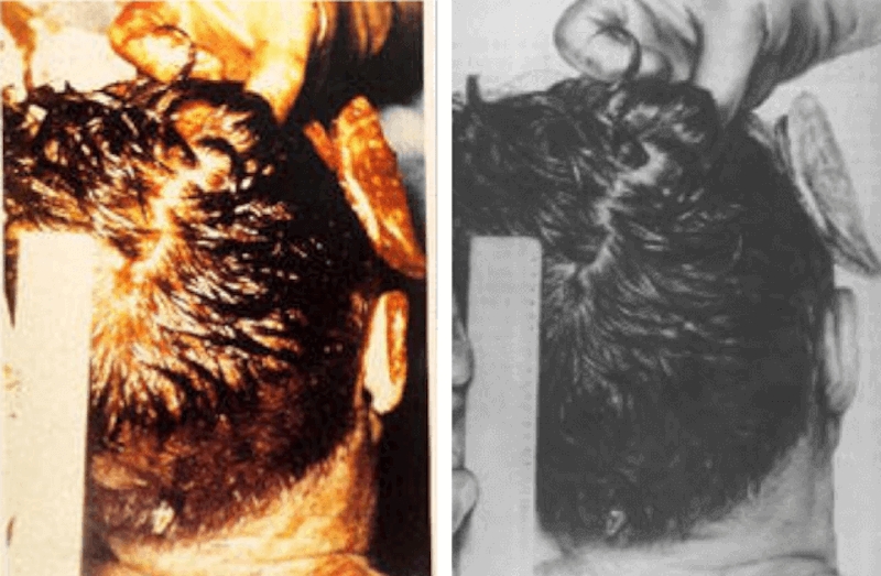
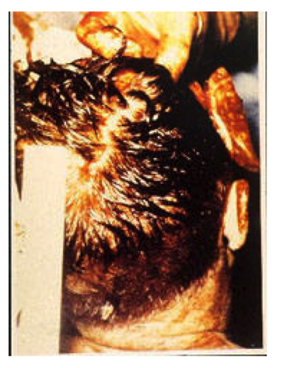
    Getting back to Dallas, in the 1990’s, some of the Parkland doctors said they never saw any defect; they said the back of the head was hidden by a curtain of gore-drenched hair that misled them into thinking a wound was under it. They also revised what they said about the brain: what they thought was cerebellum was just damaged cerebrum.

    There is a big problem with this explanation: these doctors also reported seeing damaged cerebrum, tissue which they did not mistake for cerebellum. Obviously they made a distinction between the two. And some of the exposed cerebellum was sufficiently intact to exhibit grossly visible, definable characteristics. Dr. Clark, a distinguished neurosurgeon and the most qualified of all the physicians who saw the head damage, never changed his story.

    Michael Baden, to whom Bugliosi often turned for advice, has also made good use of the hair-curtain explanation. He used it to explain how on-lookers at the autopsy could be so “wrong” about the greater defect in the skull. He even used it to explain why the pathologists were “wrong” about where the skull entrance wound was. Baden gives new meaning to the expression “pulling the wool over one’s eyes.”

    Few medical professionals would be fooled by such an explanation. Anyone who has dealt with trauma knows that even the least serious little wound in the highly vascularized scalp can cause a great blood bath. Even brain injuries can look worse than they are. Doctors and nurses always look under the mess for its source.

    Another source of the controversy: an object on the skull X-ray (frontal view), presumed to be a bullet fragment. The pathologists, the acting radiologist, and other autopsy witnesses described the largest fragment as just a sliver, shaped like a matchstick, located in the front of the head, right behind the right eye. They confirmed its location in the brain, and extracted it.

    The frontal X-ray shows something quite different: a shiny round object with the same diameter as the Carcano bullet, imbedded in the rear of the head. It shows through the eye socket, as obvious as a candle in a pumpkin. And all skull X-rays show the new location of the entrance, four inches higher. (Army experiments on skulls performed in 1964, after the autopsy report was written, showed that the lower entrance resulted in an exit that was also too low. A reason to relocate the entrance?)

    Below you will find a few specimens that reflect Bugliosi’s attempts to deal with these controversies. There are many more that I have not reported for lack of time.

    Autopsy Protocol

    Cerebellum

    Specimen 8:

    But although the autopsy report notes that “the major portion” of the right cerebrum was “exuding” from the large defect on the right side of the president’s head, there isn’t one word in the report indicating that any part of the cerebellum was missing or even lacerated. 148 (Bugliosi, p. 404)

    Specimen 9:

    It bears repeating that the autopsy report only mentioned damage to the cerebrum, not the cerebellum. (Bugliosi, p. 405)

    Specimen 10:

    Dr. Boswell, in response to Parkland doctor Kemp Clark’s claiming to have seen “exposedä cerebellar tissue,” told Dr. Gary Aguilar, “He was wrong.† The right side of the cerebrum was so fragmented.† I think what he saw and misinterpreted as cerebellum was that.” (Bugliosi, p. 405)

    Significant omission: What Bugliosi does not report is that there is not one word, one way or the other, on the appearance of the cerebellum in the main Autopsy Report or in the Supplemental Autopsy Report, where a description of the organ belonged, under the heading “Gross Description of the Brain.” (A significant omission from the autopsy protocol itself, and from Bugliosi’s description of it.)

    Another significant omission: Bugliosi does not report that in the section on the Microscopic specimens, the cerebellum (item “f. From the right cerebellar cortex”) is indeed mentioned as having “significant abnormalities … directly related to the recent trauma.” The entire quote:

    “Multiple sections from representative sections are essentially similar and show extensive disruption of brain tissue with associated hemorrhage. In none of the sections examined are there significant abnormalities other than those directly related to the recent trauma.” (CE 391, page 2, ARRB MD4)

    It is not likely the typist mistook “cerebrum” for “cerebellum.” Individual parts of the cerebrum were listed: the right parietal lobe, the right frontal lobe, the left fronto-parietal cortex — all parts of the cerebrum. The pathologists clearly described both types of brain tissue.

    It is standard to mention all normal parts of an organ adjacent to the abnormal parts, and the exclusion of the cerebellum from the Gross Description of the Brain, and its inclusion in the Microscopic Examination, is intriguing indeed.

    Occiput

    Specimen 11:

    Cerebellum certainly wouldn’t likely have been expelled from any defect in the right front of the president’s head, where the Warren Commission and the autopsy surgeons concluded the exit wound was. (Bugliosi, p.405)

    Specimen 12:

    Baden: “But, clearly from the autopsy X-rays and photographs and the observations of the autopsy surgeons, the exit wound and defect was not in the occipital area. There was no defect or wound to the rear of Kennedy’s head other than the entrance wound in the upper right part of his head.” (Bugliosi, p.408)

    As a matter of fact, the autopsy surgeons said the great defect was chiefly in the parietal area but “extended somewhat into the temporal and occipital regions.” (Autopsy Protocol, p.3) (Emphasis mine.) (And do not confuse the location of the defect with that of the exit.)

    Cerebellum “Mistaken” for Cerebrum

    Specimen 13:

    Dr. Jenkins wrote that “the cerebellum had protruded from the [head] wound … ” However, Jenkins changed his mind after seeing autopsy photographs in 1988, telling author Gerald Posner that “the photos showed the President’s brain was crenelated from the trauma, and it resembled cerebellum, but it was not cerebellar tissue.” (Bugliosi, p.405)

    Specimen 14:

    [Quoting Dr. Carrico] “Looking at the shredded pieces of brain on the gurney, it looked like some of it had the characteristics of cerebellum, which kind of has a wavy surface. But because these brain pieces were shredded, this could easily have led to confusion as to whether it was all cerebrum – which has broader bands across the surface – or some cerebellum.” (Bugliosi, p. 405)

    As Bugliosi reports, several other Parkland doctors revised their statements, but I repeat: there is a big problem with this explanation. These doctors also reported seeing damaged cerebrum, tissue which they did not mistake for cerebellum. Obviously they made a distinction between the two. Some of the exposed cerebellum was sufficiently intact to exhibit grossly visible, definable characteristics. (And it is strange that Bugliosi gives credence to anything said by Marion T. Jenkins, considering this doctor’s ability to confabulate. For details, please see my essay, “The Wandering Wounds,” at http://www.assassinationweb.com/cranrev.htm.

    The Great Hair Curtain

    Hair Hides Wound from Parkland?

    Specimen 15:

    [W]hat is the explanation for several of the other Parkland doctors erroneously thinking that the large exit wound was to the right rear of the President’s head as opposed to the right frontal region, where all the medical and scientific evidence proved it to be? Dr. Michael Baden … has what I believe to be the answer …”The head exit wound was not in the parietal-occipital area, as the Parkland doctors said. They were wrong … That’s why we have autopsies, photographs, and X-rays to determine things like this. Since the thick growth of hair on Kennedy’s head hadn’t been shaved at Parkland, there’s no way for the doctors to have seen the margins of the wound in the skin of the scalp. All they saw was blood and brain tissue adhering to the hair. And that may have been mostly in the occipital area because he was lying on his back and gravity would push his hair, blood, and brain tissue backward … (Bugliosi, pp 407-408) (Emphases his.)

    Bugliosi quotes several Parkland doctors who now say the wound was obscured by hair, “confirming” Baden’s explanation. But how could Bugliosi accept this without question even though he has shown he is familiar with testimony that contradicts it – that these doctors looked beneath the hair, and saw a defect in bone? Doctors and nurses always look under the mess for its source. Among the following quotes, notice all the references to bone:

    “[A] large wound beginning in the right occiput extending into the parietal region. Much of the skull appeared gone.” (17 WCH 10) “This was a large, gaping wound in the right posterior part, with cerebral and cerebellar tissue being damaged and exposed.” (6 WCH 20) “The loss the right occipital and probably part of the right parietal lobes would have been of specific importance. (6 WCH 26). William Kemp Clark

    “The wound … was a large gaping wound, located in the right occipitoparietal area. . . . about 5 to 7 cm. in size, more or less circular, with avulsions of the calvarium and scalp tissue.” (6 WCH 6) Carrico

    “It seemed to me that in the right occipitalparietal area that there was a large defect. There appeared to be bone loss and brain loss in this area.” (6 WCH 71) Peters

    “There was a great laceration on the right side of the head (temporal and occipital), causing a great defect.” (17 WCH, CE 392) “I really think part of the cerebellum, as I recognized it, was herniated from the wound.” (6 WCH 48) Jenkins

    “I noted a large avulsive wound of the right parietal occipital area, in which both scalp and portions of skull were absent, and there was severe laceration of underlying brain tissue.” (3 WCH 371) Perry

    “[T]he parietal bone was protruded up through the scalp and seemed to be fractured almost along its right posterior half, as well as some of the occipital bone being fractured in its lateral half, and this sprung open the bones that I mentioned in such a way that you could actually look down into the skull cavity itself and see that probably a third or so, at least, of the brain tissue, posterior cerebral tissue and some of the cerebellar tissue had been blasted out. (6 WCH 33) McClelland

    Hair Hides Wound from Autopsy Onlookers?

    Specimen 16:

    Baden said that Kennedy’s head wasn’t even shaved of its hair at the time of the autopsy, and hence, any observations by onlookers of the autopsy, as opposed, he said, to the autopsy surgeons themselves, who were working directly with the president’s head) would likely have been skewed. (Bugliosi, p.408)

    A small hole revealed by shaving the scalp is probably the one thing observers at a distance would not be able to appreciate. But these onlookers observed the scalp being reflected back to show the damage in the actual bone. Some described the brain being removed, and made other very specific observations that were based on a view of naked bone. (These witness statements have been reported so extensively by so many researchers I shall not repeat them here.) Baden apparently wishes to imply these observers saw not much more than what shows in the gory, messy photos taken before the autopsy began. Ridiculous as the comment in Specimen 15 is, Baden has topped it! See next section.

    Hair Hides Wound from Prosectors who Performed Autopsy?

    Significant omission. Bugliosi knew better than to repeat what Baden said about the four-inch discrepancy in the location of the entrance wound. In Specimen 15, Baden at least admitted that the autopsy surgeons working directly with Kennedy’s head had a better view. But you would never know it from this comment which appears in a book Baden wrote for the public:

    “Perhaps the most egregious error was the four-inch miscalculation. The head is only five inches long from crown to neck, but Humes was confused by a little piece of brain tissue that had adhered to the scalp. He placed the head wound four inches lower than it actually was, near the neck instead of the cowlick.” (Unnatural Death: Confessions of a Medical Examiner, Random House, 1989, p. 16)

    As Baden knew very well, the pathologists folded back the scalp to observe the skull directly and, they said, they looked at what was left of the hole from the inside of the skull.

    Bugliosi Blames Baden’s Co-Author

    Bugliosi admitted there were “errors” in Baden’s book, and he mentioned a few, giving the greatest space to the one concerning Pierre Finck’s background. Baden had said, falsely, that Finck had never performed an autopsy on a victim of a gunshot wound before. But Bugliosi never mentioned the two outrageous assertions from Baden’s book that I have quoted in this essay. And the excuses he makes for Baden are just not credible.

    Specimen 17:

    Baden, one of the top forensic pathologists in the nation, is an extremely busy man, and if I were to wager, he coauthored this book on the run, leaving much of the detail to his coauthor [Judith Adler Hennessee], who is not a doctor. (Bugliosi, Endnote #5, p.219)

    “Detail.” The “errors” that are the most embarrassing – the ones Bugliosi does not mention — do not concern “detail.” They are assertions concerning facts and logic treated as linchpins in proving the lone assassin theory.

    “An extremely busy man.” The chapter on the Kennedy assassination was quite small — just a few pages long — in a small book. Baden was too busy to review statements made in his name on the Crime of the Century? (Maybe he had hair in his eyes and couldn’t see the print?) “If I were to wager.” As if he had to guess. As if Baden were not available to ask directly. Considering all the direct personal contact Bugliosi had had with Baden as documented extensively in this book, you would think Bugliosi would have asked Baden himself about all of these strange statements. But, then, maybe they both were too busy.

    No Co-Author to Blame for This One

    When it came to explaining the four-inch discrepancy to Congress, Michael Baden told a different story:

    “[P]reparing the autopsy report 24 hours after the autopsy was completed and after the body had been removed, may have contributed to the more significant mistake of placing the gunshot wound of entrance 4 inches lower than it actually was. The description of the size and shape of the entry wound is correct, but the location is incorrect perhaps due to reliance on memory.” (Emphasis mine.) (1 HSCA 306)

    The location was incorrect “perhaps due to reliance on memory?” None of the congressmen questioned this. Apparently they were unaware of the notes and diagrams made during the autopsy and used in the preparation of the autopsy report. The wound, as depicted in the drawing on the autopsy descriptive sheet (ARRB MD #1), looks to be precisely at the EOP (external occipital protuberance) – low, far below another memorable landmark, the cowlick. (This interview took place before the growth of the Hair Curtain.)

    Authenticating the Skull X-rays

    Many of us are skeptical about the authenticity of the skull X-rays because what they show is just too different from what was described by the closest and most qualified witnesses. We are especially skeptical of the shiny new fragment – the perfect slice of a 6.5 Carcano bullet – that no one reported in 1964.

    David Mantik, M.D., Ph.D., a radiologist and physicist, has provided highly technical reasons for believing the X-rays are counterfeit. Bugliosi cannot deal with these concepts, and turns to wound ballistics expert Larry M. Sturdivan (BS in Physics, MS in statistics) and Dr. Chad Zimmerman for help in rebutting Mantik’s theories. What Zimmerman said about the fragment itself contradicts the opinion of the HSCA’s expert radiologist.

    Specimen 18:

    [Quoting Zimmerman] Personally, I think it may actually have been a bullet fragment that was stuck in the hair or on the skin and later fell off … I feel it is real because of the lack of film grid lines in the surrounding area, which, in my opinion, are an absolute must … in order for it to be a post-autopsy forgery. (Bugliosi, Endnotes, p.222)

    According to Gerald McDonnel, the HSCA expert radiologist, the metal fragment was imbedded on the inside of the scalp (7 HSCA 133). If McDonnel is right, it could not have been “stuck in the hair or on the skin” as Zimmerman muses.

    In any case, this does not explain why no one, including the acting radiologist at the autopsy, saw this obvious fragment on the X-ray.

    As for his opinion on what makes a forgery, what are his qualifications? Chad Zimmerman has provided Bugliosi and others with his opinions on several aspects of this case – ballistics, acoustics, neurology, radiology and photography, all promoting the lone assassin theory. He does not provide references from scholarly sources for his opinions; does this mean that he himself is a recognized scholarly source?

    With all due respect, who is Chad Zimmerman to disagree with Gerald McDonnel? He is a Doctor of Chiropractic. (Bugliosi, Endnotes, p. 327) According to his advertisements, he offers massage therapy. This case has had quite enough massage therapy.

    They Will Say Anything

    One thing is clear, if nothing else: there are people who will say anything to promote the lone assassin theory.

    It would be nice if you could just cast aside all the words and look at the images, the X-rays for instance. But here, again, you need words – the words of the people who authenticated them. Would McDonnel et al have the sophistication the spot the signs of a sophisticated forgery? Who is qualified to do that? The very people who have the expertise may be the least credible, considering their close association with the government. The relationship between Kodak and the often deceptive CIA is well established.

    Would they, too, say anything, true or not?

    How would you know?

  • Big Lie About a Small Wound


    Background

    There is no evidence, hard or soft, that supports the single bullet theory. Its defenders spend a lot of “proving” it “could have happened that way” — which is not the same as proving that it did, a distinction they don’t make. But they can’t even prove the SBT hypothetically because the sniper’s nest and the various wounds of the two men do not line up. To solve this problem, promoters of the SBT resort to chicanery. And, in desperation, they squeeze great significance out of minutiae — the small movements of Connally’s jacket and hat — events that occur all the time without the assistance of a bullet, magic or otherwise. Yet they ignore Connally’s more dramatic movements that occur too late to be associated with the same bullet that hit JFK. Worse yet, they stoop to manufacturing evidence.

    One example of manufactured evidence is the lie about Connally’s back wound. Why do supporters of the SBT say the wound was 3 centimeters long, when, in fact, it was only half as long? Why was the 1.5 centimeter wound a problem? Defenders of the theory say that if the Carcano bullet had struck sideways (as opposed to nose-on), it would have created a wound the same size as its length (3 centimeters), and such a long wound would be proof the bullet had been tumbling. If it had been tumbling, this, presumably, would be proof it had struck something else on its way to Connally’s back. The something else in this case: John F. Kennedy.

    Problem: Connally’s back wound was only as long as the wound in the back of Kennedy’s head: 1.5 centimeters — not a size that suggests the bullet was tumbling.


    clip image002

    Adaptation of a drawing demonstrating the hypothetical tumbling bullet. From John Lattimer’s book, Kennedy and Lincoln. Medical and Ballistic Comparisons of Their Assassinations, Harcourt Brace Jovanovich, 1980, page 268.

    The drawing above demonstrates the alleged behavior of the single bullet. The drawing was designed (though not executed) by John Lattimer, a urologist who has published several infomercials in medical journals promoting the lone assassin theory. What is wrong with this picture, aside from the fact that the men are too far apart? Experts assure me the Carcano bullet is much too stable to behave like this. Perforating a neck could divert the bullet, but not make it tumble to this extent in so short a time and in so short a space. (There is more on Lattimer below.)

    The wound in Connally’s back did not indicate a sideways hit any more than the wound in the back of Kennedy’s head. The latter was 1.5 x 0.6 centimeters, and the former, 1.5 x 0.8 centimeters, as documented on at least four occasions by the governor’s thoracic surgeon, Dr. Robert Shaw. (4WCH104,107; 6WCH85,86). The holes in the back of Connally’s shirt and jacket were as small as his back wound. (5WCH64) (See TABLE below.) The damage inside Connally’s chest also disproves a sideways hit. According to Shaw, the bullet created a “small tunneling wound” (7HSCA149) and he noted, “the neat way in which it stripped the rib out without doing much damage to the muscles that lay on either side of it.” (4WCH116) Shaw felt that the shape of the bullet was explained either by a “light tumbling,” or by it striking at a tangent. (6WCH95) It had to have been a tangential hit since the bullet “followed the line of declination of the fifth rib” (4WCH105), i.e., its path slanted downward.

    Connally’s back wound became 3 centimeters when it was surgically enlarged. Shaw explained that in order to clean and debride (cut away devitalized tissue) the wound, he had to enlarge it to twice its size. (6WCH88)

    Despite all this testimony, two doctors — John Lattimer and Michael Baden — found ways to make the magic bullet wound change its size to fit the magic bullet theory. And unethical “investigative journalists,” like Gerald Posner, Gus Russo and Dale Myers, have spread the lie further.

    John Lattimer Exploits a Coincidence

    Bullets and wounds are a bit like keys and locks, and that would make John Lattimer an amateur locksmith. Since a tumbling bullet did not fit into Connally’s back wound, Lattimer changed the lock.

    Some 30 years ago, Lattimer noticed an interesting coincidence: the size of the wound — after it was enlarged — was the same length as a Carcano bullet, 3 centimeters. Lattimer then published an article in which he claimed the wound had been three centimeters long — originally. (Medical Times 1974; 102 November:33-56; Kennedy and Lincoln, Harcourt Brace Jovanovich, 1980) As proof, he published Shaw’s operative report that described the wound as “three cm” — and never explained this was the size after surgical enlargement.

    Lattimer also chose to publish an uncorrected diagram that portrayed the back wound as it was after surgical enlargement (CE 679, 17WCH336) when, instead, he could have published the corrected diagram showing the actual size of the wound (Gregory Exhibit No.1, 20WCH32). See Exhibits A and B.

    On this same deceptive diagram, Lattimer pasted a snippet of testimony in which Shaw agrees the wound, as portrayed in the diagram, is correct — but, omitted from this snippet were statements that made it clear Shaw was talking about the EXIT wound. Lattimer did report that a correction had been made, but lied about its nature: “His careful diagram of the wound of entry (which he revised and initialed) showed it to be elongated in its vertical (not horizontal) axis and to be at least 3 cm in length.” (Medical Times 1974; 102 November:33-56; Kennedy and Lincoln, Harcourt Brace Jovanovich, 1980, page 266) See Exhibits B – F.

    Lattimer included Shaw’s testimony in which he agrees with Arlen Specter that the wound could have been caused by a slightly tumbling bullet — but omitted Shaw’s opinion that it could also have been caused by a tangential hit. (6WCH95) See Exhibit G. Again, it should be noted that this wound was the same length as the entrance into JFK’s head, which supporters of the lone assassin theory say was a tangential hit.

    Michael Baden Stretches the Lie Further

    Dr. Michael Baden who headed the HSCA medical panel, knew the true size of the wound and how it had been enlarged. He had also personally examined the scar on Connally’s back. This is how he described it to the panel:

    “On removing his shirt, it was readily apparent that at the site of gunshot perforation of the upper right back there is now a 1 1/8 -inch long horizontal pale well healed…” (7HSCA 143-144; 240)

    In a book Baden wrote for the public, he doubled the size of the scar:

    “According to Connally’s medical records, the bullet struck him nose first in the back and left a vertical scar. I thought the records were wrong. If it was the same magic bullet, it would have gone in sideways — with the length, not the point, first. After leaving Kennedy, it would have lost its power and became a tumbling bullet, and tumbling bullets rotate. When they finally strike, they strike edgewise. I needed to examine Connally…

    “He removed his shirt. There it was — a two-inch long sideways entrance scar in his back. He had not been shot by a second shooter but by the same flattened bullet that went through Kennedy.” (Unnatural Death: Confessions of a Medical Examiner, Random House 1989, p.20) See Exhibit H.

    Apparently Baden realized that if the original wound had been three centimeters (1.2 inches), then, after its surgical enlargement, the scar would have been even bigger. It was a calculated lie. This is only one of several examples of Michael Baden’s deceptions.

    Gerald Posner, Gus Russo, and Dale Myers Perpetuate the Lie

    Two of these “investigative reporters,” Posner and Russo, never quoted primary sources on the wound, choosing instead to accept the fraudulent claims of Lattimer and Baden. (Gerald Posner. Case Closed. Random House, pp 336, 479; Gus Russo. Live by the Sword: The Secret War Against Castro and the Death of JFK. Bancroft Press, 1998, p.297).

    The third “investigative reporter,” Dale Myers, mimicked Lattimer’s technique of taking testimony out of context. On his website, he wrote: “According to Dr. Robert Shaw’s operative record, the entrance wound in JBC’s body was ‘just lateral to the right [shoulder blade] close [to] the [armpit] yet has passed through the latysmus [latissimus] dorsi muscle…the wound of entrance was approximately [1.2 inches] in its longest diameter.’” (7HSCA142) But Myers omitted what followed immediately after the quote above: “The rear entrance wound was NOT [emphasis mine] 3 centimeters [1.2 inches] (in diameter) as indicated in one of the operative notes. It was a puncture-type wound, as if a bullet had struck the body at slight declination [i.e., not at a right angle]. The wound was actually approximately 1.5 centimeters in diameter. The ragged edges of the wound were surgically cut away, effectively enlarging it to approximately 3 centimeters.” (7HSCA143)

    Conclusion

    Five people, including the head of the second biggest investigation into the medical evidence, Michael Baden, MD, have demonstrated great faith in the public’s inability or unwillingness to make a simple comparison between what they say, and what is a matter of public record.

    Copyright © 2004, by Milicent Cranor


    table

    exhibit a

    exhibit b

    exhibit c

    exhibit d

    exhibit e

    exhibit f

    exhibit g

    exhibit h 19

    exhibit h 20

  • Michael Baden’s Deceptions


    From Probe, Volume 4, Number 4 (May-June 1997)


    (Click here if your browser is having trouble loading the above.)


  • The Magician’s Tools


    From the November-December, 1995 issue (Vol. 3 No. 1) of Probe


    “The most important tools of the magician are diversion and timing.”
    ~ Anonymous Victorian conman, inspiring his class of beginning pickpockets with the secrets of a loftier trade.


    Seriously Out of Synch

    Three shells, three shots. When was shot #2? According to the government, Kennedy and Connally, hit by shot #2, are wounded five seconds before the head shot. Yet, shots #2 and #3 have been called “close together.” Shots fired five seconds apart might be called “close together” but never a “flurry.”

    The vast majority of witneses said they heard only one shot before the two or more “close together” shots. Many of these witnesses were not only explicit about what they meant by close together (“simultaneous”), they were also clear about when the flurry occurred – during the time the head exploded. Before that, they heard only one shot, apparently the one that struck Kennedy in the throat. What happened in between?

    By all accounts, the first shot was fired before the limousine disappeared behind the Stemmons Sign (from Zapruder’s viewpoint). Most of us believe he reacts to the throat wound at this time. Kennedy emerges from behind the sign and, as others have noted, he goes forward a few inches – violently. This little movement, obscured by his suddenly rising arms, may indicate a separate shot to the back. Soon after, Connally is hit. Kellerman said he heard only one shot, then no more until the time of the “flurry.” Clint Hill said he heard one shot before the “double sound” he associates with the head wound, and nothing in between. (In the Altgens photo, the equivalent of Z-255, Hill has not yet reacted.) Samuel A. Kinney heard one shot, then a second, and “the second shot was fired and I observed hair flying from the right side of his head.” I’m assuming this second shot was a head shot because of the similarity between his testimony and George Hickey’s: “I heard a loud report…[then] I heard two reports which … appeared to me completely different in sound than the first report and were in such rapid succession there seemed to be no time element between them. The first shot of the second two seemed as if it missed because the hair on the right side of his head flew forward and there didn’t seem to be any impact against his head. The last shot seemed to hit his head…” Again, a second shot associated with disturbing Kennedy’s head. Rather than “hair” flying forward, I think what they saw was the bone flap that appears seven frames before the acknowledged head shot on the Nix film (I describe corroborating evidence for this in “The Magic Skull,” The Fourth Decade, July, 1995).

    Some witnesses seemed unaware of even the first shot. Mary Moorman, for example. She took two Polaroid photos of Kennedy. By the time she took the second picture (the head shot), at least two shots had already been fired. Yet, she said she heard a shot for the first time as she took this second picture. Charles Brehm is another witness who seemed unaware of the shots at first. As noted by David Lifton, Brehm is clapping his hands as the car passes him, a time when at least two shots had been fired.

    An Apparent Contradiction

    Governor Connally said he did not hear the shot that hit him. Neither, apparently, did Mrs. Connally. She said she heard a shot, turned around, saw Kennedy in distress, and kept her eyes on him until her husband was hit.

    “I never again looked in the back seat of the car after my husband was shot. My concern was for him…” The strange thing is, except for a quick glance at Connally as he changes posture, Mrs. Connally continuously stares at Kennedy – for two full seconds of surviving film after her husband has been hit. Another oddity: Governor Connally said he cried out, “Oh, no,no,no,” after he was hit. Mrs. Connally claimed he said it before he was hit.

    I think they were both right.

    Mrs Connally could have meant that before she heard the second shot, Connally cried out. What she really heard was probably the first of the flurry that struck Kennedy’s head.

    Evidence of Subsonic Bullets

    Robert Shaw, MD described the hole through Connally as a “small tunneling wound,” noting the “neat way in which it stripped the rib out without doing much damage to the muscles that lay on either side of it.” Kennedy’s back wound was apparently even less. A supersonic bullet would have done much more damage. One thing that would render a supersonic bullet subsonic is a silencer.

    Perception of Gunfire

    There is usually variation among witnesses in the perception of anything, especially gunfire. Variation in hearing ability, attention span, the location of witnesses compared with the location of the marksmen, all could create apparent inconsistencies. A shot fired from 100 yards away is 20 decibels softer than one fired 10 yards away [20xlogarithm (base 10) of the ratio of the distances], all other factors being equal. Furthermore, loud noises are deafening, just as bright lights are blinding, temporarily. The pupil contracts in bright sunlight and takes time to adjust to a dark room, and muscles supporting the eardrum contract in self defense following a loud noise, making the ear deaf to quieter sounds. Thus, two shots could sound like one, depending on the witness’s location. And one shot can sound like two if the location is conducive to echo. Three shots from the same place, fired five seconds apart, would produce either three echoes, or no echo. That sound follows impact isn’t a factor here because of the distances involved. How to know which sounds go with which wounds? Will Greer said the last two shots were fired “simultaneously,” and that Connally moved at that time. Hit earlier, Connally was probably only flinching, or reacting to yet another shot. Greer’s assumption is probably “witness confusion,” a real phenomenon, but it could mean any testimony that does not agree with your theory.

    Prying Apart the Last Two Shots

    “Close together” could mean “closer together,” if the time between shots #1 and #2 could be lengthened, which may be why some claim shot #1 came as early as Z-160. But another solution to the problem is this hypothetical statement:

    “You only heard an echo. Look at this map. There were only three shots, and witnesses say the car was clear over here near this sign, when Connally fell. That was shot #2. And the car was down here several feet away when the head opened up. That was shot #3. You couldn’t have heard a flurry, the car didn’t move that fast. We even have it on film.”

    Zapruder was on his perch well before the motorcade arrived, and he could have been removed “for security reasons.” And surely they knew in advance that people film such motorcades. Did they want a record of it, one they could edit? One that would record the “second” shot? Bang……..Bang…….Bang?

    BANG…………..BANG-BANG

    The following people heard only one shot before the flurry, which occurred at the time the head exploded:


    Jack Bell (AP): “in quick succession” (NYT 11/23/63, p.5)
    George Hickey: “in rapid succession…no time element between” (18H762)
    Roy Kellerman: “flurry…plane breaking the sound barrier…bang, bang”; (2H76)
    Clinton Hill: “The second shot had “an echo…double sound” (2H144)
    Mary Woodward: “The second two shots were immediate…as if one were an echo of the other… with the second and third shot…I saw the head explode” (A∓E,II )
    Will Greer: “simultaneously” (2H118)
    Glenn Bennett: “A second shot followed immediately and hit the right rear high of the President’s head.” (18H760)
    Rufus Youngblood: “in rapid succession.” Rufus Youngblood (Robert MacNeil’s The Way We Were, 1988, Carroll ∓ Graf)
    Warren Taylor: “in the instant that my left foot touched the ground, I heard two more bangs” (CE1024)
    Seymour Weitzman: “simultaneous” (7H106)
    Linda Kay Wills: “two real fast bullets together” (7H498)
    Lee Bowers: Rapped his knuckles on a table showing the near simultaneity of the last two. (Mark Lane 1966 Tape)
    Junior Jarman: “third shot was fired right behind the second” (3H204)
    Carolyn Walther: “almost at the same time” (C.E.2086)
    Toney Henderson: “in rapid succession” (C.E.208)
    Mrs. Lyndon Johnson: “in rapid succession.” (H565)


    Mr. and Mrs. William Newman who stood on the curb slightly to the left of Zapruder describe the opposite pattern, BANG-BANG, nothing, then BANG. This may be related to location.

    Conclusion

    The purpose of silently creating wounds between audible shots #1 and #2 may not have been to disprove the closeness of shot #2 to shot #3, but one thing seems certain: what did the majority of witnesses hear when Connally was shot? Nothing.

  • The FBI’s Fib about the Mannlicher Carcano


    From the July-August, 1995 issue (Vol. 2 No. 5) of Probe


    FBI firearms expert Robert Frazier, testifying before the Warren Commission, described the results of tests by FBI marksmen with Oswald’s Mannlicher Carcano at 15, 25 and 100 yards. Their shots consistently landed close to each other, within an area “the size of a dime,” but not close to the target, demonstrating the rifle’s precision, but lack of accuracy due to the misalignment of the telescopic sight. (Accuracy and precision have separate meanings in ballistics.) Their results:

    At 15 yards:

    2.5 to 4 inches too high;

    1 inch too far to the right

    At 25 yards:

    4 to 5 inches too high;

    1 to 2 inches too far to the right

    At 100 yards:

    2.5 to 5 inches too high;

    2 to 5 inches too far to the right

    If the bullet struck 2.5 – 4 inches too high at 15 yards, how could it be a mere 2.5 to 5 inches too high at 100 yards?

    Deviation is in direct proportion to the distance of the gun from the target. Earlier in his testimony, in a very different context, Frazier made an offhand remark that illustrates this principle: He said he fired three shots at 25 yards with “approximately a 3-inch spread…the equivalent of a 12-inch spread at a hundred yards.” Twelve inches, not 5 inches?

    Would bullet drop (effect of gravity) compensate for the rifle’s poor vertical alignment at 100 yards? I got a precise answer from the editor of a leading ballistics publication who, because of the “sensitive” subject matter, wishes to remain anonymous. Using Barnes’ Ballistics computer program, he determined that, at 120 yards, a 6.5mm, 160-grain bullet, muzzle velocity of 2,200 feet per second, would have dropped only 0.7 inches below “flat firing” level. (In a different context, Frazier claimed more bullet drop than my expert, 1.2 inches at 100 yards. Not enough to explain the results obtained.) So much for gravity explaining the disproportionately small degree of deviation at 100 yards.

    I then posed another question for his computer: if the telescopic sight of the rifle places the same bullet 3 inches above the target at 25 yards, how far above the target would the bullet strike if the rifle was zeroed in at 100 yards? He came up with 14 inches. If the bullet is 4 inches off at 25 yards, it would be 18 inches off at 100 yards. (These figures are conservative; even at 15 yards, when firing for accuracy and not speed, two of the FBI marksmen were off by 4 inches.)

    How did the FBI manage to fire “only” 5 inches too high at 100 yards (assuming they were telling the truth)? It is reasonable to conclude that, having become familiar with the gun by the time of the last series of tests, they compensated for the misalignment of the telescopic sight – and did not say so. Commissioner Eisenberg appears to have guessed it:

    “Mr. Frazier, when you were running, let’s say, the last test, could you have compensated for this defect?”

    “Yes; you could take an aiming point low and to the left and have the shots strike a predetermined point…”

     Or, was it his point that Oswald compensated for the defect? Eisenberg also appears to have known, in advance, what might solve the problem, as acknowledged:

    “[I]f the elevation crosshair was defective at the time of the assassination…and no compensation was made for this defect, how would this have interacted with the amount of lead which needed to be given to the target?”

    Frazier answered, perhaps as predicted, that no lead would have been necessary: The misaligned scope “accomplished the lead” for him. Earlier, Frazier had testified that Oswald would have had to lead (aim ahead of the target because it would have moved by the time the bullet arrived) the target by 4 to 6 inches. (If Oswald were as good a shot as claimed, would he not have aimed ahead of the target, assuming he didn’t know the sight was off?) The sight was well stabilized when received in Dallas, as shown by the shots landing so close together, but it was misaligned. Why? Frazier could not answer, but suggested it had been bumped, as evidenced by a “severe scrape on the scope tube” that occurred at some unknown time. And he said “It may be the that the mount has been bent or the crosshair ring shifted.” (Wouldn’t it be have been clear whether, if not when, the mount was bent?) Did the FBI or the Commission inquire if the scrape had been on the gun when found in the Depository? If the scrape was “severe,” wouldn’t it have been seen in Dallas? If not, the Commission could have claimed the gun was damaged in transit, and was fine at the time of the assassination. Was this basic, obvious question ever asked?

    Frazier minimized the problem, claiming it wasn’t really defective, that “only the adjusting mechanism does not have enough tolerance to bring the crosshair to the point of impact of the bullet,” simple to fix by slipping a “shim” under the sight. But, the defect is apparently inherent with that brand, and was there before the hypothetical bump. When, for his experiments, John Lattimer bought four Carcanos – “a favorite among European riflemen” – and four telescopic sights identical to Oswald’s, he found that all four needed shims, and hinted that Oswald had used one. No shim was ever found on or near Oswald’s gun.

    We may never know the truth about that gun. But we do know the FBI told what amounts to a lie. When they made the statement that, at 100 yards, the rifle’s aim was off by only 5 inches, they knew it would be understood to mean that the last series of tests was performed under the conditions of the first two tests, that is, without compensating for the misalignment of the sight. How would the public have responded to the information that, when firing the last shot, the bullet would have gone at least 14 inches above the point of aim on Kennedy’s head? The gun seems to have been more of a threat to the pigeons above. How would the public have responded to the information that the FBI rigged the last test?